Lipid nanoparticle structure, self-assembly, and endosomal escape
A mechanistic look at how ionizable lipids, PEG-lipids, cholesterol, and helper lipids cooperate to encapsulate mRNA and release it into the cytoplasm.
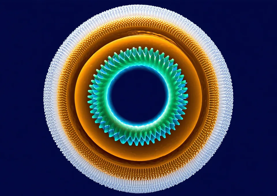
LNP cross-section: ionizable lipid core (cyan), PEG-lipid corona (teal), mRNA payload (amber), cholesterol (white dots)
The four-component LNP
Every LNP formulation is a balance of four excipients, each with a distinct function.
Ionizable Lipid
The functional core: neutral at physiological pH (7.4), protonated in the endosome (pH 5.0–6.5). Protonation drives membrane fusion and mRNA release. pKa range 6.2–6.8 is optimal.
Helper Lipid
DSPC, DOPE, or DPPC stabilize the bilayer structure and modulate membrane fluidity. DOPE's cone shape facilitates hexagonal phase transition during endosomal escape.
Cholesterol
Increases membrane rigidity, affects particle size and PDI. Regulates membrane fusion propensity. Typical range 35–45 mol%.
PEG-Lipid
Steric stabilization prevents aggregation and opsonization. PEG density (0.5–3.5 mol%) trades off serum stability against endosomal escape rate.
Why pKa 6.2–6.8 is the sweet spot
An ionizable lipid with apparent pKa < 6.0 is insufficiently protonated at endosomal pH, producing weak membrane disruption and low mRNA release. Above pKa 7.0, the lipid carries positive charge at physiological pH — which promotes non-specific protein binding and clearance by phagocytes before the particle reaches the hepatocyte.
The therapeutic window is narrow. Gendelivr's in silico pKa model predicts apparent pKa within ±0.15 units across a library of 1,200 ionizable lipid structures.
Apparent pKa and Activity
More science from Gendelivr
CRISPR-Cas9 Delivery
Why LNP-mediated mRNA CRISPR delivery outperforms viral vectors for in-vivo hepatic editing.
ReadHepatic Targeting
ApoE-mediated endocytosis and the hepatocyte targeting mechanism behind LNP organ selectivity.
ReadFormulation Engine
See how Gendelivr applies this biology to screen 10,000 formulations in silico before bench synthesis.
Explore